Utrasound Examination
UTRASOUND EXAMINATION (Aplio 500)
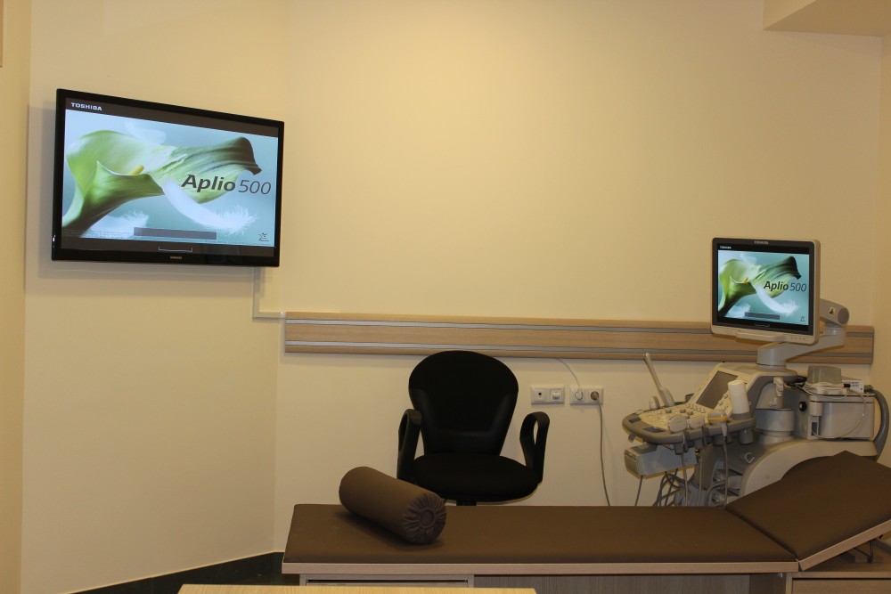
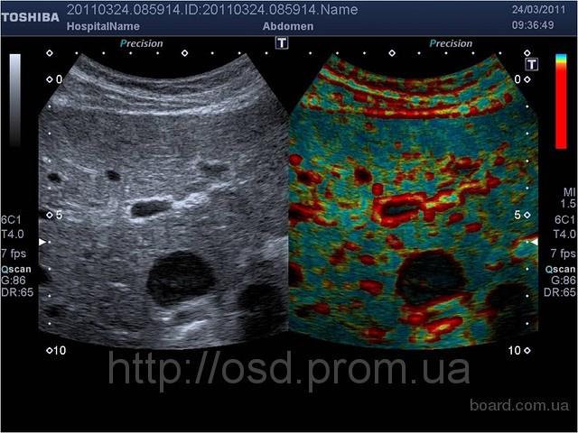
“SIRMED” medical center is equipped with TOSHIBA Aplio 500 ultrasound system, a unique 3D/4D technology in Armenia, which is one of the most innovative technologies of visualization. Through Aplipure, Aplipure+, Defferential THI, Precision Imaging modes the system provides the best visualization of tissues. Aplio 500, a Japanese premium-class ultrasound diagnostic system, is considered state-of-the-art technology of the new millennium, due to the following unique functional possibilities: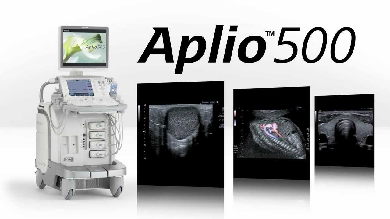
- Fly Thru mode is one of the latest 3D technologies of dimensional visualization, which opens new perspectives in diagnostic sphere. Fly Thru functional option, comparable to virtual endoscopy, provides an opportunity to “travel” virtually in the slots of hollow organs, ducts and arteries. Similarly is implemented the visualization of the mentioned organs from outer area, which is called Inversia. By this method it is possible to receive additional information about tumors and invasive growth of dimensional formations.
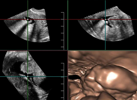
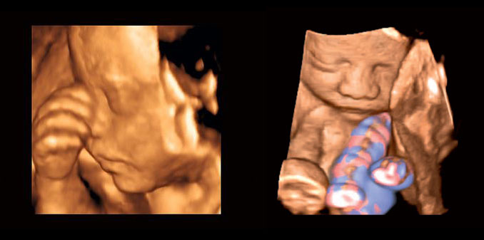
- Superficial 3D reconstruction and 4D LIVE-real time 4D scanning via the homonymous transducer is broadly used in midwifery, especially for visualization of fetus.
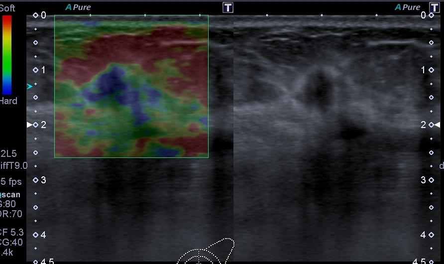
- ELASTO Q-ultrasound elastography or sonoelastography is one of the newest and most promising methods in ultrasound examination sphere, which is based on the peculiarity of elasticity of different tissues. Via Aplio 500 the difference of tissues is estimated not only by color, but also by quantitative parameters. This method provides an opportunity to make the distinguishing diagnosis between benign and malign tumors.
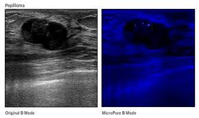
- MicroPure mode provides an opportunity to identify even the smallest microcalcinates in thyroid gland, mammary gland, kidneys and other organs.
- Panoramic mode provides an opportunity to proceed with new examination up to several meters long, while keeping the previous image on the monitor, which increases the scope of information and assists the doctor`s work.
- The examination through CEUS- Ultrasound contrast amplification is applicable with all types of transducers.
- For transthoracic examination of cardio-vascular system the ultrasound examination, by means of the software, provides best-quality image. The flexible M-mode, the complex programs of measurements and the module of reports create comfortable conditions for stress echocardiography. In particular, mention-worthy is the Wall Motion Tracking (WMT) technology, the application of which is simple and which provides an opportunity to take the measurements with different velocities, determine the type of deformation, and promptly estimate the heart and cardio-vascular system from the structural and functional viewpoints. The IMT has a significant importance in diagnosing the vascular diseases, through which it is possible automatically to measure the intima media of vessel wall, simplifying and accelerating the examination process.
PRICE LIST
| THE NAME OF SERVICE |
COST /AMD/ |
| Ultrasound examination of abdominal and small pelvic organs |
15.000 |
| Ultrasound examination of abdominal organs |
10.000 |
| Ultrasound examination of female pelvic organs by (vaginal) sensor by vaginal sensor |
10.000 |
| Ultrasound examination of female pelvic organs by (vaginal) sensor |
8.000 |
| Foliculometry |
13.000 |
| Ultrasound examination of fetus, fetometry (3D/4D) |
10.000 |
| Ultrasound examination of urinary |
7.000 |
| Fetal Doppler examination |
15.000 |
| Ultrasound examination of thyroid and parathyroid glands |
8.000 |
| Elastography ultrasound examination of thyroid glands and determination of microcalcinates` existence (in one node) |
10.000 |
| Elastography ultrasound examination of thyroid glands and determination of microcalcinates` existence (in two and more nodes) |
15.000 |
| Ultrasound examination of salivary glands |
10.000 |
| Ultrasound examination of lymph nodes (one district examination) |
6.000 |
| Ultrasound examination of lymph nodes (all districts examination) |
15.000 |
| Complex ultrasound examination of cervical organs (thymoidglands, parathymoid glands, salivary glands, lymph nodes) |
15.000 |
| Ultrasound examination of mammary glands |
10.000 |
| Elastography ultrasound examination of mammary glands and determination of microcalcinates` existence |
15.000 |
| Ultrasound examination of soft tissues |
6.000 |
| Ultrasound examination of soft tissues with elastography |
10.000 |
| Ultrasound examination of soft tissues with elastography by 18L*7 matrix sensor |
6.000 |
| Ultrasound examination of joints |
13.000 |
| Ultrasound examination of joints by elastography and determination of microcalcinates |
15.000 |
| Ultrasound examination of minor bones |
15.000 |
| Complex sonography of abdominal organs, small pelvic organs, thyroid glands and mammary glands |
25.000 |
| Ultrasound examination of testicle |
10.000 |
| Ultrasound examination of testicle and urinary |
15.000 |
| Duplex examination of testicle |
15.000 |
| Neurosonography |
10.000 |
| Transcranial neurosonography |
15.000 |
| Ultrasound examination of thymus glands |
10.000 |
| Duplex examination of magistral arteries Arteries and veins of superior /upper/ extremity Arteries and veins of lower extremities Duplex scanning of brachiocephalic arteries |
15.000 15.000 20.000 |
| Provision of electronic copy of ultrasound examinations` results |
3.000 |
| Liver elastography and duplex scanning |
30.000 |
| Liver elastography |
20.000 |
| Nerve conduction study and electromyography (electroneuromyography) |
25.000 |
| Nerve conduction study and electromyography (electroneuromyography), repeat examination (within 40 days) |
15.000 |
| Echocardiogram |
15.000 |
| Echocardiogram (by provision of electronic copy) |
25.000 |
| Echocardiogram by Wall Motion Tracking technology |
15.000 |
| Stress echocardiogram |
20.000 |
| Electrocardiogram |
3.000 |
| ECG monitoring 24 hours |
15.000 |
| ECG monitoring 48 hours |
25.000 |
| Blood pressure monitoring |
10.000 |
| Tredmil test |
15.000 |
|
For foreign citizens the prices are doubled |
|
| Zephyr Life-monitoring | 3 days |
45.000 |
7 days |
70.000 |
14 days |
100.000 |
20 days |
150.000 |
30 days |
200.000 |
Consultation of invited as a professional (cosultant) |
20.000 |
For foreign sitizens the prices are doubled |
||
How to arrive to the ultrasound examination
Ultrasound examination of abdominal organs is performed in hungry condition (it is desirable not to eat, drink tea or coffee for 6-8 hours before examination). If the examination is made in the afternoon or diabetic patients should undergo the examination, the consumption of not sweet tea with a piece of bread rings is allowed. One day before the examination all kind of food should be excluded from ration that may cause of flatulence (black bread, milk, peas, fresh fruits and vegetables).
Ultrasound examination of small pelvic organs (uterus, ovaries, prostate) is performed under full bladder conditions. For this, 1.5-2 hours prior the examination 1 litre of still liquid drink (water, juice or tea) should be consumed. It is desirable to implement the ultrasound examination of mammary glands after menstrual cycle. For examination of thyroid and parathyroid glands, salivary glands, lymph nodes, soft tissues, arteries of upper and lower extremities, brain arteries, elastography and echocardiography no special preparations are needed.
